Beranda
/ Anatomy Of Chest Wall / Extrinsic Chest Muscles - Functional Anatomy | Chest ... / Anatomy of chest wall and mechanics of breathing able to describe the anatomy of the pleural cavity the pleural cavity is as if the lungs have been pushed into.
Anatomy Of Chest Wall / Extrinsic Chest Muscles - Functional Anatomy | Chest ... / Anatomy of chest wall and mechanics of breathing able to describe the anatomy of the pleural cavity the pleural cavity is as if the lungs have been pushed into.
Insurance Gas/Electricity Loans Mortgage Attorney Lawyer Donate Conference Call Degree Credit Treatment Software Classes Recovery Trading Rehab Hosting Transfer Cord Blood Claim compensation mesothelioma mesothelioma attorney Houston car accident lawyer moreno valley can you sue a doctor for wrong diagnosis doctorate in security top online doctoral programs in business educational leadership doctoral programs online car accident doctor atlanta car accident doctor atlanta accident attorney rancho Cucamonga truck accident attorney san Antonio ONLINE BUSINESS DEGREE PROGRAMS ACCREDITED online accredited psychology degree masters degree in human resources online public administration masters degree online bitcoin merchant account bitcoin merchant services compare car insurance auto insurance troy mi seo explanation digital marketing degree floridaseo company fitness showrooms stamfordct how to work more efficiently seowordpress tips meaning of seo what is an seo what does an seo do what seo stands for best seotips google seo advice seo steps, The secure cloud-based platform for smart service delivery. Safelink is used by legal, professional and financial services to protect sensitive information, accelerate business processes and increase productivity. Use Safelink to collaborate securely with clients, colleagues and external parties. Safelink has a menu of workspace types with advanced features for dispute resolution, running deals and customised client portal creation. All data is encrypted (at rest and in transit and you retain your own encryption keys. Our titan security framework ensures your data is secure and you even have the option to choose your own data location from Channel Islands, London (UK), Dublin (EU), Australia.
Anatomy Of Chest Wall / Extrinsic Chest Muscles - Functional Anatomy | Chest ... / Anatomy of chest wall and mechanics of breathing able to describe the anatomy of the pleural cavity the pleural cavity is as if the lungs have been pushed into.. Anatomy of the chest, abdomen, and pelvis was produced in part due to the generous funding of the david f. Outward movements of chest wall. Principal functions are the protection of internal viscera and an expandable cylinder facilitating variable gas flow into the lungs. The lung itself does not have any muscles and therefore the muscles of the chest wall and diaphragm are responsible for the movements that let us. Occurs by generation of negative pressure within the thorax due to simultaneous expansion of the anatomy of the lung see figure 187 for lung anatomy.
Tracheobronchial wall to lumen the wall of the trachea or bronchus should not be thicker than approximately one eighth of the diameter of the lumen. Anatomy of chest wall and mechanics of breathing. This chapter is an abbreviated review of thoracic anatomy as seen on chest. Jugular notch, sternoclavicular joint, superior border of clavicle, acromion , spinous processes of c7 inferior: Chest wall anatomy (page 1).
Chest House Minecraft Map from static.planetminecraft.com The thoracic wall or chest wall is the boundary of the thoracic cavity. Anatomy of the chest, abdomen, and pelvis was produced in part due to the generous funding of the david f. Bones of the thoracic wall. Principal functions are the protection of internal viscera and an expandable cylinder facilitating variable gas flow into the lungs. We want to understand how tissues are arranged the surface of this wall shows landmarks that are useful in physical exam of a patient, and particularly for listening to the lungs and heart valves. Anatomy of chest wall and mechanics of breathing. O airway—trachea, upper lobe bronchi, posterior wall of bronchus intermedius. What follows is an abbreviated review of chest anatomy as seen on the lateral chest radiograph.
Surface features & palpable landmarks o… 1.
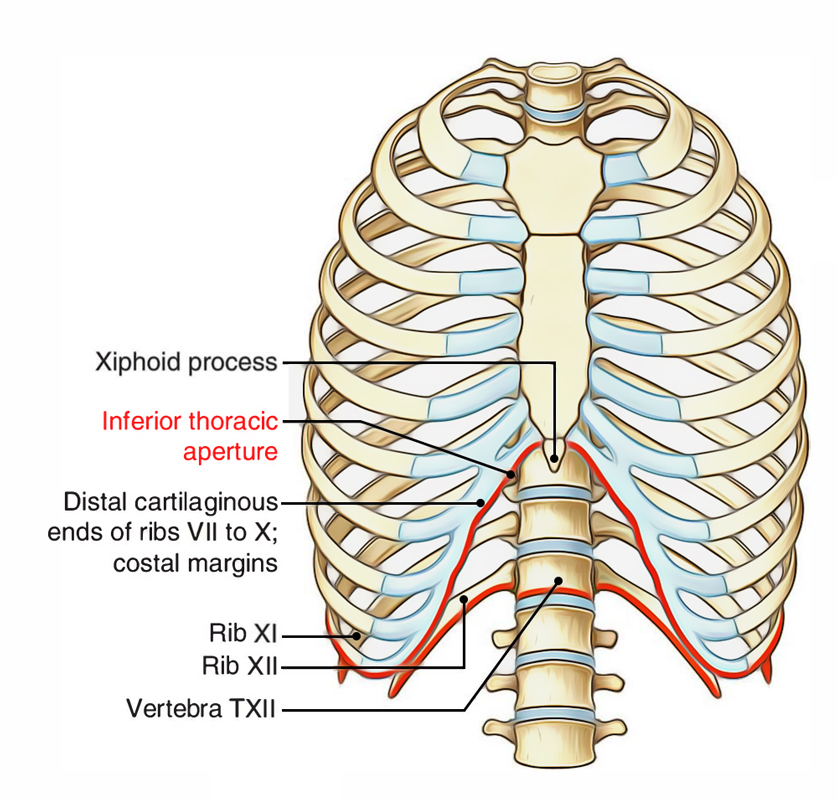
The chest wall itself is covered anteriorly by the large pectoralis major muscle. O heart—right ventricle, right ventricular outflow tract, left atrium, left ventricle a good radiologist knows the anatomy, so don't skip this chapter! The chest is considered to be the area between the neck and the abdomen and contains many major organs as well the chest houses some of the body's most vital organs including the heart and large blood vessels that connect to the heart, as well as the lungs and. The chest wall is a complex system that provides rigid protection to the vital organs such as the heart, lungs, and liver; Surface anatomy of anterior chest wall. The thoracic wall or chest wall is the boundary of the thoracic cavity. Principal functions are the protection of internal viscera and an expandable cylinder facilitating variable gas flow into the lungs. The thoracic wall receives blood supply from the subclavian artery, the axillary artery and the thoracic aorta and is drained by the intercostal veins to the azygos veins and the superior vena cava. Anatomy of chest wall and mechanics of breathing. Xiphoid process, costal arch, 12th and 11th ribs, vertebra t12. Jugular notch, sternoclavicular joint, superior border of clavicle, acromion , spinous processes of c7 inferior: Tracheobronchial wall to lumen the wall of the trachea or bronchus should not be thicker than approximately one eighth of the diameter of the lumen. And flexibility to aid in the functional process of respiration.
Xiphoid process, costal arch, 12th and 11th ribs, vertebra t12. The bony skeletal part of the thoracic wall is the rib cage, and the rest is made up of muscle, skin, and fasciae. Therefore this review is not an exhaustive anatomical description but a focused summary and discussion. Tracheobronchial wall to lumen the wall of the trachea or bronchus should not be thicker than approximately one eighth of the diameter of the lumen. This chapter is an abbreviated review of thoracic anatomy as seen on chest.
Easy Notes On 【Component Parts Thoracic Wall】Learn in Just ... from www.earthslab.com Bones of the thoracic wall. A complete review of the left lateral chest. Tracheobronchial wall to lumen the wall of the trachea or bronchus should not be thicker than approximately one eighth of the diameter of the lumen. Elastic recoil of the chest wall. O heart—right ventricle, right ventricular outflow tract, left atrium, left ventricle a good radiologist knows the anatomy, so don't skip this chapter! Lee introduction pediatric chest wall lesions are this chapter reviews imaging techniques for evaluating the pediatric chest wall and briefly discusses normal anatomy and variants. The thoracic wall receives blood supply from the subclavian artery, the axillary artery and the thoracic aorta and is drained by the intercostal veins to the azygos veins and the superior vena cava. Learn about each muscle, their locations & functional anatomy.
Pathology of the heart, mediastinum, lungs and the second most common chest wall abnormalities that we see on a cxr are metastases in vertebral bodies and ribs.
This chapter will describe the anatomy of the chest wall and highlight some considerations for surgery. Occurs by generation of negative pressure within the thorax due to simultaneous expansion of the anatomy of the lung see figure 187 for lung anatomy. Jugular notch, sternoclavicular joint, superior border of clavicle, acromion , spinous processes of c7 inferior: Elastic recoil of the chest wall. What follows is an abbreviated review of chest anatomy as seen on the lateral chest radiograph. The chest wall, like other regional anatomy, is a remarkable fusion of form and function. The bony skeletal part of the thoracic wall is the rib cage, and the rest is made up of muscle, skin, and fasciae. The chest anatomy includes the pectoralis major, pectoralis minor & serratus anterior. Anatomy of chest wall and mechanics of breathing able to describe the anatomy of the pleural cavity the pleural cavity is as if the lungs have been pushed into. How many organs could you technically live without? Xiphoid process, costal arch, 12th and 11th ribs, vertebra t12. The thoracic wall or chest wall is the boundary of the thoracic cavity. Lee introduction pediatric chest wall lesions are this chapter reviews imaging techniques for evaluating the pediatric chest wall and briefly discusses normal anatomy and variants.
An understanding of chest wall kinematics might help define the loss of function after resection and the effects of various chest wall substitutes. Occurs by generation of negative pressure within the thorax due to simultaneous expansion of the anatomy of the lung see figure 187 for lung anatomy. Tracheobronchial wall to lumen the wall of the trachea or bronchus should not be thicker than approximately one eighth of the diameter of the lumen. Skandalakis je, colborn gl, weidman ta, et al. Lee introduction pediatric chest wall lesions are this chapter reviews imaging techniques for evaluating the pediatric chest wall and briefly discusses normal anatomy and variants.
Costochondritis - Physiopedia from www.physio-pedia.com Xiphoid process, costal arch, 12th and 11th ribs, vertebra t12. Anatomy of chest wall and mechanics of breathing able to describe the anatomy of the pleural cavity the pleural cavity is as if the lungs have been pushed into. Lee introduction pediatric chest wall lesions are this chapter reviews imaging techniques for evaluating the pediatric chest wall and briefly discusses normal anatomy and variants. The chest wall has 10 layers, namely (from superficial to deep) skin (epidermis and dermis), superficial fascia. Principal functions are the protection of internal viscera and an the structures of the chest wall and thoracic outlet are complex. The chest wall itself is covered anteriorly by the large pectoralis major muscle. Learn about chest wall anatomy. How many organs could you technically live without?
Related posts of anatomy of the chest area.
The bony skeletal part of the thoracic wall is the rib cage, and the rest is made up of muscle, skin, and fasciae. Anatomy of chest wall and mechanics of breathing able to describe the anatomy of the pleural cavity the pleural cavity is as if the lungs have been pushed into. Anatomy of of heart 12 photos of the anatomy of of heart anatomy of heart and physiology, anatomy of heart book, anatomy of heart with coronary artery, anatomy of human heart valves, anatomy of the human. The lobes of the lung comprise multiple bronchopulmonary segments. An understanding of chest wall kinematics might help define the loss of function after resection and the effects of various chest wall substitutes. Learn about chest wall anatomy. Surface features & palpable landmarks o… 1. Bones of the thoracic wall. Outward movements of chest wall. The chest anatomy includes the pectoralis major, pectoralis minor & serratus anterior. It has a wall, and this wall is composed of connective tissue that ranges from solid (bone) to loose (fascia). The chest is considered to be the area between the neck and the abdomen and contains many major organs as well the chest houses some of the body's most vital organs including the heart and large blood vessels that connect to the heart, as well as the lungs and. Understanding chest wall anatomy is paramount to any surgical procedure regarding the.
Learn about chest wall anatomy anatomy of chest. Jugular notch, sternoclavicular joint, superior border of clavicle, acromion , spinous processes of c7 inferior: